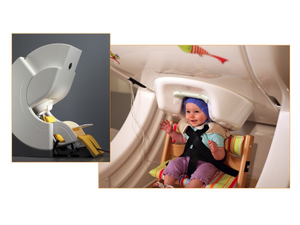


We know that experiences influence brain development. But teasing apart the details of this intricate dance of experience and biology has been a challenge. Now, cutting-edge tools are providing scientists with a new window into the developing brain. Brain imaging techniques like magnetoencephalography reveal what parts of the brain are active. Magnetoencephalography, or MEG, is able to detect the location and intensity of brain activity. As a child interacts with the world around them, we get a glimpse into their brain.
This brain imaging tool is silent, non-invasive and harmless. MEG can’t read minds – scientists aren’t able to tell what we are thinking or feeling using this tool. Instead, MEG can tell us what parts of our brain are active as we do certain tasks, like reading or listening to foreign language sounds. MEG detects the weak magnetic field produced by the symphony of neurons talking to each other deep inside the brain. This type of information helps scientists understand how different parts of the brain work together.
-
- Axon
- output fiber of a neuron
- Cell body
- the neuron's processing center
- Dendrites
- input fibers of a neuron
- Magnetoencephalography (MEG)
- a non-invasive brain imaging technique used to determine which regions of the brain are active
- Neurons
- cells located in the brain and throughout the body that are specialized to communicate messages
- Pruning
- the process of removing excess synapses
- Sensitive period
- a time in development when the brain is especially ready to learn a skill
- Synapse
- connection point between neurons